Cone Beam CT (CBCT)
Cone Beam CT სკანირება
Dental Cone Beam CT scans (CBCT Scans) provide high resolution, 3D volumetric images, used in Dental Implant Planning, Orthodontics and Maxillofacial surgery. allowing for more accurate analysis of bone structure and dental/tooth orientation. The accuracy of a CBCT scan is comparable to medical CT scans but uses a much lower radiation dose but with the added advantage of greater accuracy of bone structures and dental/tooth orientation. Your patient sits in a chair, as opposed to lying in a tunnel, and the time for a dental CBCT scan is just twenty seconds.
Whereas typical dental X-rays focus on a small area to produce flat, 2D image, CBCT scan images enable a more complete and accurate 3D images - allowing you to more effectively plan patient surgical and dental treatments. Individual patient Cone Beam CT images are also digitally stored for ease of access in the future, and for sharing with colleagues or patients.
Cone Beam CT სკანირება მრავალ სიტუაციაში გამოიყენება და სარგებელს გაძლევთ შემდეგ სფეროებში:
დენტალური იმპლანტები:
CBCT სკანირების მაღალი ხარისხი საშუალებას გაძლევთ უფრო ზუსტად დაგეგმოთ და განათავსოთ იმპლანტები — დიაგნოსტიკის საწყისი ეტაპიდან მკურნალობასა და ოპერაციის შემდგომ გამოკვლევებამდე.
ორთოდონტია:
CBCT ორთოდონტიაში იძლევა მკაფიო და ზუსტ სურათებს ბრეკეტების განთავსებისთვის, ასევე კბილების ურთიერთმდებარეობისა და ასიმეტრიის შეფასებას.
ჩაჭედილი კბილები:
A CBCT scan can highlighted whether a patient has impacted teeth and accurately determine their precise position in relation to adjacent teeth and associated roots, and proximity to important structures such as nerve canals, sinus walls and cortical borders.
პაციენტის სასუნთქი გზების შეფასება:
Restricted airways are susceptible to collapse but are not always easy to pinpoint. Cone beam CT scan of a patients’ airway allows you to reconsider your treatment plan if a problem is found.
ტემპორომანდიბულარული სახსარი (TMJ):
Temporomandibular joint CBCT (TMJ CBCT) is not only cost-effective but allows complete TMJ analysis and diagnosis of multiple TMJ conditions, providing clear images of joint spacing and any dysfunction of condyles and surrounding structures.
ანალიზი:
Cone Beam CT სკანირების ანალიზი გამოიყენება პაციენტის შესაძლო ან გაუთვალისწინებელი პრობლემების აღმოსაჩენად, ძვლისა და ყბის დეფორმაციების ზუსტი გაზომვის წყალობით. ეს საშუალებას გაძლევთ შეაფასოთ ძვლის დაზიანებები და ყბის ცვლილებები, აღმოაჩინოთ კისტები, სიმსივნეები და დაავადებები.
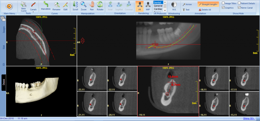
CBCT-ის სამუშაო მაგალითი:
Click on the image below to open up a working example of a Cone Beam CT scan visualised using the PACS Cloud Viewer
სწრაფი შეკითხვა
დაგვირეკეთ დღესვე+44 (0)20 7487 5717